
What do you do if you want to look inside a biological cell, hoping to see objects that are well beyond the diffraction limit of a microscope – roughly 200nm? One solution is to use the Nobel-prize-winning technique of “super-resolved fluorescence microscopy”, which involves tagging samples with fluorescent markers.
In its basic form, however, super-resolution microscopy only produces 2D images, meaning it does not produce any “depth” information in 3D. That’s where Double Helix Optics, a small firm in Boulder, Colorado, comes in.
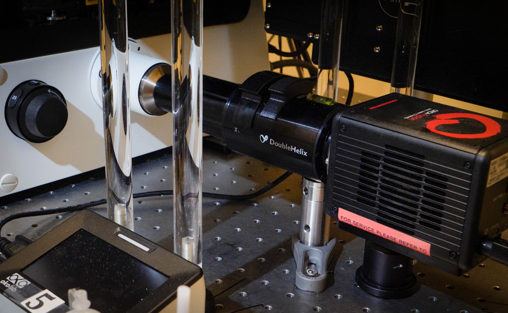
As I found out on a recent visit, this start-up company has developed a small optical device, called a SPINDLE, that can be bolted on to a standard wide-field optical microscope, allowing you to use “out-of-focus” light to generate 3D images at super resolution.
In this short video, Ronald Zimmerman – Double Helix’s director of sales and product management – introduces the basic principles of the device and outlines possible applications, including studying the motion of individual viruses. The video was filmed just before the great global coronavirus lock-down, which makes that particular application potentially more valuable and useful than ever.
Find out more about SPINDLE on the Double Helix Optics website.